Cardioception: The Bidirectional Connection Between the Heart and the Brain
"The heart is more than a mechanical pump; this vital organ can function as an independent 'small brain,' even influencing our behaviors, emotions, and decision-making."
"In the world of science, new discoveries sometimes challenge long-held beliefs. The present article examines the bidirectional relationship between the heart and the brain, demonstrating that the heart is not merely a blood-pumping organ but an independent information-processing center capable of influencing cognition, emotions, and even human personality. By integrating findings from neuroscience, Qur’anic studies, and medicine, this research opens new horizons in understanding the complex interaction between the body and the mind. Has the time not come to revisit the traditional definition of the heart?"
Abstract
Numerous verses in the Holy Qur’an refer to the heart as an instrument for reasoning, contemplation, deliberation, and understanding. In many instances, it is mentioned alongside the eyes and ears, suggesting that the heart, like these sensory organs, possesses perceptual capabilities and serves as a gateway for information entering the body. Since the 17th century, when William Harvey’s discovery of the circulatory system established the heart’s role solely as a blood pump, most commentators—avoiding conflict with scientific knowledge—have interpreted these Qur’anic references within the realm of moral philosophy, disregarding the definition of the heart as a physiological organ with material properties. Despite this caution, the Qur’an’s relatively explicit descriptions of the heart as a tool for reasoning or perception—similar to the eyes and ears—cannot be overlooked, particularly where it assigns the heart a location within the thoracic cavity.
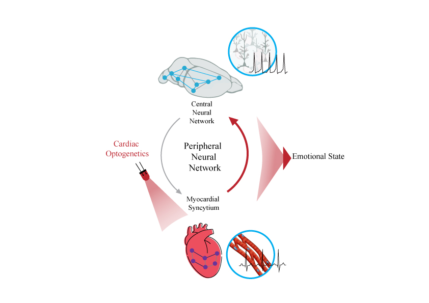
On the other hand, in the field of empirical sciences, since the discovery of the heart’s intrinsic nervous system (the “heart’s small brain”) in 1991, various studies have reported the heart’s relative autonomy and its high degree of independence from the brain. This has provided an opportunity to revisit questions such as: Are emotions the cause of the brain’s responses or their consequence? Does anxiety lead to an increased heart rate, or does the heart drive anxiety-related behaviors? Different methodologies have been proposed to address these questions, the most significant of which are introduced in this observational study.
Keywords: Cardiac interception, shared heart-brain disorders, the heart’s small brain.
1. Introduction and Research Background
The heart constitutes one of the most pivotal concepts in the epistemological framework of the Holy Qur’an. Within Islamic intellectual heritage, the Qur’an attributes numerous cognitive functions—such as contemplation (tafakkur), reasoning (ta‘aqqul), and understanding (tafaqquh)—to the heart in 132 verses, positioning it as a sensory organ alongside instruments like the ears and eyes. To avoid conflating the Qur’anic concept of the heart with empirical sciences, many exegetes have confined its meaning to the realm of ethics, advocating a purely spiritual interpretation while disregarding its physiological dimensions as a material organ.
Despite this caution, the Qur’an’s relatively explicit references to the heart as an instrument of reasoning or perception—analogous to the eyes and ears—cannot be overlooked. For instance, Qur’an 22:46 locates the heart within the thoracic cavity, while Qur’an 33:4 (“Allah has not made for any man two hearts within his body”) underscores its corporeal nature. Similarly, Imam Ali (AS) explicitly affirms the heart’s physicality in Sermon 108 of Nahj al-Balagha, enumerating its spiritual attributes.
Historically, the identification of the brain as the center of cognition was gradual. Ancient Greek philosophers like Aristotle and Plato held divergent views: Aristotle regarded the heart as the seat of perception and bodily control, dismissing the brain as a mere blood-cooling organ, whereas Plato emphasized the brain’s role in intelligence. Aristotle’s theory, bolstered by its alignment with religious, philosophical, and popular beliefs of the era, dominated for centuries due to insufficient anatomical knowledge. This paradigm was challenged in two phases: first, by Galen (2nd century CE), whose dissections revealed the brain’s role in cognition via the nervous system; and second, during the Renaissance, when William Harvey’s discovery of blood circulation (17th century) definitively refuted the heart’s cognitive role. By the 19th–20th centuries, neuroimaging technologies erased all doubt about the brain’s supremacy.
However, the 1991 discovery of the heart’s intrinsic nervous system (“the heart’s small brain”) revealed its relative autonomy from the brain, reigniting questions such as: Are emotions the cause or consequence of neural responses? Does anxiety elevate heart rate, or does the heart drive anxiety-related behaviors? Emerging methodologies, particularly optogenetics, now enable causal studies of cardiac dynamics on brain function. Understanding heart-brain interactions is thus critical to deciphering the heart’s role in decision-making and behavioral modulation.
2. Methodology
2-1. Theological and Qur’anic Methodology
The Qur’an mentions the heart in 132 verses, linking it to cognitive behaviors like reasoning (‘aql), jurisprudential insight (fiqh), and reflection (tadabbur). Among these, 40 verses reference reasoning—one explicitly tied to the heart—while deep contemplation (yafqahūn) appears in 20 verses (7 associating it with the heart, 1 with the chest). Reflection (tadabbur) is cited in 4 verses, one attributing it to the heart. These functions unequivocally implicate the heart in cognition, either as a primary agent or a key facilitator.
This study analyzes these verses alongside authoritative translations and exegeses. Despite Imam Ali’s explicit acknowledgment of the heart’s physicality in Nahj al-Balagha, modern exegetes have largely avoided this interpretation to prevent conflict with empirical science. We thus examine whether a physiological reading of these verses contradicts established scientific knowledge.
2-2. Empirical Methodology
A comprehensive review was conducted on studies investigating Cardioception (cardiac interception), methods to disrupt this pathway, and links between heart-brain diseases. Emphasis was placed on landmark studies that transformed scientific understanding. Results are categorized into:
Clinical studies on the correlation between heart and brain diseases (e.g., stress cardiomyopathy, neurocardiology).
3. Key Studies Confirming Bidirectional Heart-Brain Interactions and Future Research Directions
3-1. Heart Interception
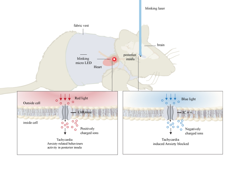
3-1-1. Optogenetic Cardioception Anxiety and profound emotional responses are deeply linked to the autonomic system, particularly the heart and brain. While the brain’s effects on heart rate are well-documented, the causal role of the heart in driving behavioral states—especially anxiety—remains underexplored. A recent study employed advanced optogenetic tools to investigate how artificial heart rate alterations influence psychological states and behavior. This pioneering study, which induced tachycardia in freely moving mice via external optical stimulation independent of brain activity, provides the first experimental evidence of a causal heart-brain-behavior axis. Its findings, published in top-tier journals like Nature and Science, resolve centuries of debate by demonstrating the heart’s active role in modulating behavior [1].
Methodology: The study used optogenetics to artificially manipulate cardiac rhythms in mice and assess anxiety-like behaviors. Key tools included:
Wearable micro-LEDs for noninvasive cardiac stimulation.
Brain activity imaging and optogenetic inhibition of specific neural circuits.
Behavioral assays (elevated plus maze, open field, risk-reward tasks).
Technical Details:
Red Light for Cardiac Stimulation:
Viral Vectors: Adenovirus (AAV9) delivered the light-sensitive protein ChRmine to cardiomyocytes under the cardiac-specific mTNT promoter.
Stimulation: 590 nm red light from a chest-mounted micro-LED induced artificial tachycardia (up to 900 bpm).
Blue Light for Neural Inhibition:
Viral Vectors: AAVs delivered *iC++*, a hyperpolarizing opsin, to neurons in the posterior insular cortex.
Insular Cortex Role: The posterior insula mediated cardiac signal processing; its optogenetic inhibition reduced anxiety despite tachycardia [3].
Cardio-Behavioral Defense: Heart rate shifts (bradycardia/tachycardia) were context-dependent and regulated by periaqueductal gray neurons [4].
Heart-to-Brain Signaling: Cardiac signals directly altered neural activity and behavior without environmental triggers [5].
Implications: This study confirms the heart’s active role in emotional states, with cardiac signals routed via the insula to modulate behavior. Optogenetics offers noninvasive, cell-type-specific control over heart-brain interactions, though translational challenges remain
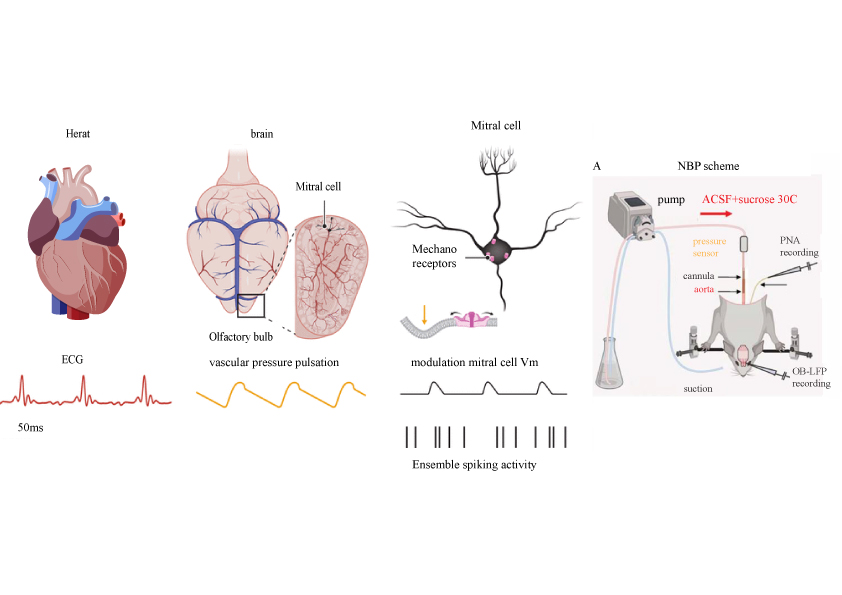
3-1-2. Mechanical Cardioception: Heartbeat Perception Speed The cardiovascular system influences cognition and emotion via mechanical signals. Recent studies highlight how heartbeat-induced pressure waves interact with brain oscillations (e.g., theta, LFP) through Piezo2 mechanosensitive channels, enabling faster signal transmission than synaptic pathways [6–9].
Methods:
Semi-Intact Models: Isolated olfactory bulbs perfused with artificial pulsatile flow to exclude synaptic/respiratory confounds.
Piezo2 Modulation: Channel blockade (GsMTx4) and optogenetic inhibition.
EEG-ECG Synchronization: Revealed heartbeat-locked neuronal spiking in mitral cells (20 ms latency).
Heartbeat-Brainwave Coupling: 15% of olfactory mitral cells were heartbeat-sensitive, firing synchronously with cardiac cycles.
Mechanical Signal Speed: Piezo2-mediated transmission was orders of magnitude faster than synaptic pathways.
This rapid mechanosensory pathway prioritizes cardiac feedback for real-time cognitive-emotional regulation, offering novel targets for cardio-cerebral disorders.
3-1-3. Mechanosensory Channels in Cardioception Heartbeats generate vascular pressure waves detected by Piezo2 channels, which modulate cognition and emotion [10].
Heart-Brain Axis:
Top-Down: CNS regulates heart rate via sympathetic/parasympathetic pathways.
Bottom-Up: Mechanical cardiac signals are transduced by Piezo2 in mitral cells, influencing neuronal activity.
Clinical Applications:
Psychiatric Disorders: Piezo2 dysfunction may underlie autism/schizophrenia sensory processing deficits.
BCIs: Mechanosensory channels could enhance brain-computer interfaces.
Cardio-Psychiatric Therapies: Real-time pressure monitoring may predict mental health risks.
Challenges: Signal variability, respiratory interference, and pathway complexity necessitate further research.
3-1-4. Heart Rate Variability (HRV) and Brain Function This study examined how reduced HRV (via TGAC8 overexpression) alters neural activity and behavior [7, 11].
Predictive Coding Framework: The brain uses cardiac timing to allocate resources between interoception and exteroception.
3-1-7. Cardiac Sensory Neurons in Cognition The heart’s 40,000 sensory neurons (its “little brain”) project to prefrontal/insular/limbic regions, influencing risk assessment and decision-making [14].
Translational Potential: Biofeedback therapies targeting cardiac signals may improve cognitive performance under stress.
3-1-8. Cardioception as a Component of Interoception The heart’s intrinsic nervous system (sensory/motor/interneurons) communicates bidirectionally with the brain via vagal/spinal pathways, modulating reflexes (e.g., baroreflex) and emotional states [15].
Future Directions:
Genetic/anatomical mapping of cardio-cerebral circuits.
Long-term cognitive effects of cardiac interventions.
Conclusion
These studies redefine the heart as an active participant in cognition and emotion, with mechanosensory, optogenetic, and HRV-based mechanisms offering novel therapeutic avenues. Future work should prioritize human translational models and real-world clinical applications.
3-2. The Correlation Between Heart and Brain Diseases
3-2-1. Effects of Heart Transplantation on Behavior, Personality, and Cognition
Traditionally viewed as a mechanical pump for blood circulation [16], the heart is now recognized as an organ capable of influencing cognition, perception, and even human behavior. The concept of cardioception—the internal sensing of cardiac activity—has opened new avenues in neuroscience and medicine. This section examines post-heart transplant changes in personality, cognitive impairments, and their impact on patient survival. Heart transplantation, a life-saving intervention for end-stage failure, is often associated with shifts in personality, behavior, and cognition [17–19].
Personality and Behavioral Shifts Post-Transplantation: Reports indicate that many recipients experience altered food preferences, emotional responses, and even self-identity perceptions. The cellular memory hypothesis posits that epigenetic, DNA, or RNA mechanisms in donor hearts may drive these changes [20, 21]. Anecdotal cases describe recipients adopting traits resembling their donors, though empirical evidence remains limited [22].
Post-Transplant Cognitive Dysfunction: Cognitive impairments—reduced information processing speed, memory deficits, and executive dysfunction—affect 30–60% of recipients [23]. These deficits diminish quality of life, treatment adherence, and survival rates, particularly in older patients with comorbidities like diabetes or low left ventricular ejection fraction (LVEF) [24].
Beyond a Pump: A Paradigm Shift in Cardiology: Current transplant protocols prioritize the heart’s mechanical role, overlooking its neurocognitive significance. To improve outcomes, three reforms are proposed:
Clinician Education: Enhances awareness of the heart’s role in neural-cognitive networks [11, 24].
Conclusion: Post-transplant behavioral and cognitive changes underscore the heart’s role in higher-order brain functions. A holistic approach integrating cardiac and neural health is essential to optimize patient care.
3-2-2. Disrupted Heart-Brain Synchrony in Major Depressive Disorder (MDD)
A study investigating heart-brain coupling in MDD patients analyzed theta wave-brain and cardiac cycle (systole/diastole) synchrony using simultaneous EEG-ECG in 90 MDD patients and 44 controls [25].
MDD Patients: Markedly reduced synchrony, especially during diastole, correlated with symptom severity (e.g., hopelessness). Antidepressants partially restored this synchrony.
Implications: The Jaccard Index (JI), introduced here, may serve as a diagnostic tool for heart-brain dyssynchrony, highlighting bidirectional heart-brain communication in mental health.
3-2-3. Structural Heart-Brain Links
A UK Biobank MRI study (N=40,000) analyzed 82 cardiovascular and 458 brain traits, identifying 80 shared genetic loci [26].
Mechanisms and Impacts:
Cardiac Dysfunction (e.g., atrial fibrillation): Reduces cerebral blood flow, damaging white matter and elevating dementia risk.
Shared Genetics: Ventricular wall thickness correlated with cortical thinning, implicating cardiac traits in stroke, schizophrenia, and depression.
Limitations: While MRI confirmed phenotypic-genetic links, the study overlooked potential cognitive roles of the heart beyond hemodynamics.
3-2-4. Stress and Cardiovascular Health
Research on stress-induced neural activity (SNA) and cardiovascular outcomes [27, 28] reveals:
SNA-Mortality Link: SNA predicts all-cause mortality but weakly correlates with major adverse cardiac events (MACE).
Arrhythmia→Depression: Cardiac uncertainty and lifestyle disruptions fuel depressive cycles.
Therapeutic Insight: Dual-target therapies (e.g., SSRIs + beta-blockers) may break this vicious cycle.
“The heart’s independent neural networks and signaling capacity—affecting decision-making, anxiety, and sensory perception—align with Quranic references to cardiac ‘reasoning.’ This demands a neuroscientific redefinition of the heart’s role beyond mere physiology.”
4. Conclusion and Future Research Directions
The present study, through comprehensive empirical and theoretical analysis, elucidates the indispensable role of the heart in bidirectional interactions with the central nervous system. The findings robustly demonstrate that the heart functions not merely as a mechanical pump, but rather as an active signaling organ that transmits independent and influential neural inputs to the brain via specialized networks, modulating cognitive processes, emotional regulation, and even complex behaviors. Converging evidence from optogenetic studies, neuroimaging, and electrophysiological research collectively confirms that the heart-brain axis operates through sophisticated mechanisms that transcend traditional neurocardiac models.
From a clinical perspective, these results indicate that cardiovascular disorders may directly or indirectly impair cerebral functioning—and vice versa—underscoring the necessity for integrated diagnostic and therapeutic approaches to related pathologies. Furthermore, the alignment of certain findings with concepts articulated in religious texts creates fertile ground for deeper interdisciplinary investigations bridging neuroscience, medicine, and Islamic studies.
Future Research Directions:
Molecular/Cellular Mechanisms: Deciphering the role of specific ion channels (e.g., Piezo2) in cardiac-to-neural signal transmission.
Translational Models: Developing advanced animal and human models to precisely map cardiac-induced cognitive-behavioral effects.
Clinical Applications: Innovating therapeutic interventions for anxiety and depressive disorders via heart-brain axis modulation.
Cross-Cultural Studies: Exploring conceptual synergies between contemporary scientific discoveries and philosophical-theological perspectives on the heart.
In summary, this research necessitates a paradigm shift in mainstream neuroscience and medical frameworks, redefining the heart not as a passive organ, but as a dynamic and influential component within the intricate “mind-body” network. Future advancements in this field may catalyze transformative insights into the embodiment of cognition and emotion.
References:
1. Rodriguez-Rozada, S., S. Frantz, and P. Tovote, Cardiac optogenetics: regulating brain states via the heart. Signal Transduction and Targeted Therapy, 2023. 8(1): p. 324.
2. Hsueh, B., et al., Cardiogenic control of affective behavioural state. Nature, 2023. 615(7951): p. 292-299.
3. Martini, E., The cardiac origin of anxiety. Nature Cardiovascular Research, 2023. 2(4): p. 339-339.
4. Signoret-Genest, J., et al., Integrated cardio-behavioral responses to threat define defensive states. Nature Neuroscience, 2023. 26(3): p. 447-457.
5. Couderc, Y. and A. Beyeler, How an anxious heart talks to the brain. 2023, Nature Publishing Group UK London.
6. Hamill, O.P., Arterial pulses link heart-brain oscillations. Science, 2024. 383(6682): p. 482-483.
7. Whalley, K., Olfactory neurons can feel the (heart) beat. Nature Reviews Neuroscience, 2024. 25(4): p. 210-210.
8. Jammal Salameh, L., et al., Blood pressure pulsations modulate central neuronal activity via mechanosensitive ion channels. Science, 2024. 383(6682): p. eadk8511.
9. Zhao, Y., et al., Rapid coupling between vasculature and neurons through mechanosensitive channels in the olfactory lobe. Frontiers in Human Neuroscience, 2024. 18: p. 1435859.
10. Xu, S., et al., Heart-brain connection: How can heartbeats shape our minds? Matter, 2024. 7(5): p. 1684-1687.
11. Khalsa, S.S., Rhythms of the Heart, Echoes in the Brain: Exploring Interoception. 2023, American College of Cardiology Foundation Washington DC. p. 2236-2239.
12. Klein, A.S., et al., Fear balance is maintained by bodily feedback to the insular cortex in mice. Science, 2021. 374(6570): p. 1010-1015.
13. Al, E., et al., Heart–brain interactions shape somatosensory perception and evoked potentials. Proceedings of the National Academy of Sciences, 2020. 117(19): p. 10575-10584.
14. Tendulkar, M., et al., Clinical potential of sensory neurites in the heart and their role in decision-making. Frontiers in Neuroscience, 2024. 17: p. 1308232.
15. Lovelace, J.W., J. Ma, and V. Augustine, Defining cardioception: Heart-brain crosstalk. Neuron, 2024.
16. Sirri, L., et al., Manifestations of health anxiety in patients with heart transplant. Heart & Lung, 2020. 49(4): p. 364-369.
17. Mauthner, O.E., et al., Heart transplants: Identity disruption, bodily integrity and interconnectedness. Health:, 2015. 19(6): p. 578-594.
18. Jones, B.M., et al., Psychological adjustment after cardiac transplantation. Medical Journal of Australia, 1988. 149(3): p. 118-122.
19. Anthony, S.J., et al., The heart as a transplanted organ: Unspoken struggles of personal identity among adolescent recipients. Canadian journal of cardiology, 2019. 35(1): p. 96-99.
20. Liester, M.B., Personality changes following heart transplantation: The role of cellular memory. Medical hypotheses, 2020. 135: p. 109468.
21. Carter, B., et al., Personality Changes Associated with Organ Transplants. Transplantology, 2024. 5(1): p. 12-26.
22. Bürker, B.S., et al., Cognitive function among long‐term survivors of heart transplantation. Clinical Transplantation, 2017. 31(12): p. e13143.
23. Xiong, T., et al., The impact of postoperative cognitive impairment on mid‐term survival after heart transplantation. Clinical Transplantation, 2023. 37(3): p. e14870.
24. Qin, Q., et al., Postoperative cognitive dysfunction in heart transplantation recipients. Clinical Transplantation, 2024. 38(5): p. e15337.
25. Zhou, H., et al., Brain–heart interaction disruption in major depressive disorder: disturbed rhythm modulation of the cardiac cycle on brain transient theta bursts. European Archives of Psychiatry and Clinical Neuroscience, 2024. 274(3): p. 595-607.
26. Zhao, B., et al., Heart-brain connections: Phenotypic and genetic insights from magnetic resonance images. Science, 2023. 380(6648): p. abn6598.
27. Fayad, Z.A. and D. O’Connor, Unveiling the heart’s silent whisperer: study of stress and the brain–heart connection in Europe. 2024, Oxford University Press UK. p. 1631-1633.
28. Mikail, N., et al., Imaging of the brain–heart axis: prognostic value in a European setting. European heart journal, 2024. 45(18): p. 1613-1630.
29. Liao, G.-z., et al., Exploring the heart-brain and brain-heart axes: Insights from a bidirectional Mendelian randomization study on brain cortical structure and cardiovascular disease. Neurobiology of Disease, 2024. 200: p. 106636.
30. Fang, S. and W. Zhang, Heart–Brain Axis: A Narrative Review of the Interaction between Depression and Arrhythmia. Biomedicines, 2024. 12(8): p. 1719.